You are here :
Science Fair 2021
Science Fairs are one of the ways the EUTOPIA alliance chose to display the variety and the quality of its PhD and post-doctoral research. Due to the pandemic, the first EUTOPIA Science Fair will be organised as a virtual event—a Research Posters exhibition in EUTOPIA’s website—starting 15 February 2021.
The Science Fair intends to show the research developed by young and confirmed researchers, finishing a PhD or in a post-doctoral position, from all over the EUTOPIA Alliance.
What?
- A virtual exhibition displayed on EUTOPIA’s website of 60 Research Posters. The Science Fair is open to all research areas, without restrictions.
- An enriched digital publication, with all the posters, the abstracts and the videos.
- A roundtable on Scientific Communication the 25 February 2021.
- A real exhibition of the printed posters in the campuses of the EUTOPIA’s six universities, when the COVID-19 pandemic will allow it.
Visit the EUTOPIA Science Fair:
- 1. The World of Matter: Physics
-
- Dominique Frizon de Lamotte et al., CY Cergy Paris Université
-
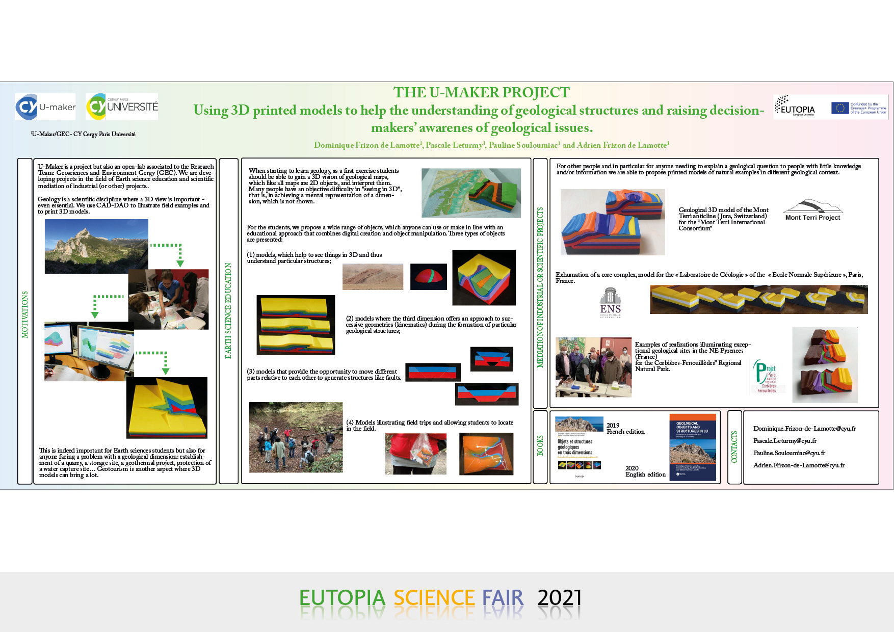
The U-MAKER Project: Using 3D printed models to help the understanding of geological structures and raising decision-makers’ awareness of geological issues
DOMINIQUE FRIZON DE LAMOTTE, PASCALE LETURMY, PAULINE SOULOUMIAC AND ADRIEN FRIZON DE LAMOTTE
U-Maker/GEC- CY Cergy Paris Université
U-MAKER is a project but also an open-lab associated to the Research Team: Geosciences and Environment Gergy (GEC). We are developing projects in the field of Earth science education and scientific mediation of industrial (or other) projects.
Geology is a scientific discipline where a 3D view is important – even essential. When starting to learn geology, as a first exercise students should be able to gain a 3D vision of geological maps, which like all maps are 2D objects, and interpret them. Many people have an objective difficulty in “seeing in 3D”, that is, in achieving a mental representation of a dimension, which is not shown.
This is indeed important for Earth sciences students but also for anyone facing a problem with a geological dimension: establishment of a quarry, a storage site, a geothermal project, protection of a water capture site… Geotourism is another aspect where 3D models can bring a lot.
For the students, we propose a wide range of objects, which anyone can use or make in line with an educational approach that combines digital creation and object manipulation. In fact, our computer-designed prototypes are saved in a format from which they can be printed in 3D. Three types of objects are presented:
1. Models, which help to see things in 3D and thus understand particular structures;
2. Models where the third dimension offers an approach to successive geometries (kinematics) during the formation of particular geological structures;
3. Models that provide the opportunity to move different parts relative to each other to generate structures like faults;
4. Models illustrating field trips and allowing students to locate in the field.
For other people and in particular for anyone needing to explain a geological question to people with little knowledge and/or information, we are able to propose printed models of natural examples in different geological context. - Guy Trambly de Laissardière et al., CY Cergy Paris Université
-
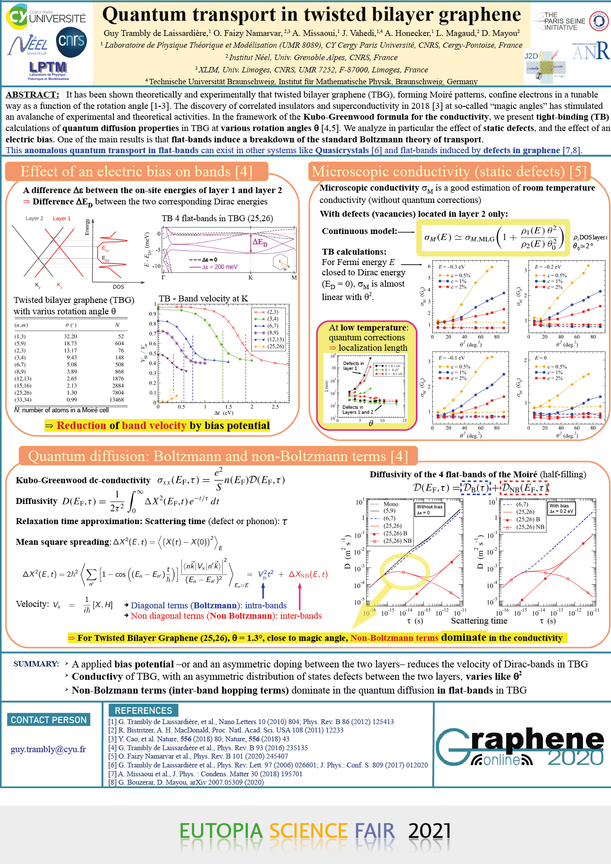
Quantum transport in twisted bilayer graphene
- Karol Hricovini et al., CY Cergy Paris Université
-
Topological states at the InBi(100) surface
KAROL HRICOVINI (1), JÁN MINÁR (2), CHRISTINE RICHTER (1), OLIVIER HECKMANN (1), JEAN-MICHEL MARIOT (3), JANUSZ SADOWSKI (4), JÜRGEN BRAUN (5), HUBERT EBERT (5), JONATHAN DENLINGER (6), IVANA VOBORNIK (7), JUN FUJIIK (7), PAVOL ŠUTTA (2), MARTIN GMITRA (8) AND AND LAURENT NICOLAÏ (2)
(1) Laboratoire de Physique des Matériaux et des Surfaces, CY Cergy Paris Université, France; karol.hricovini@cyu.fr
(2) University of West-Bohemia, Plzeň, Czech Republic
(3) Laboratoire de Chimie Physique-Matière et Rayonnement, Sorbonne Université, Paris, France
(4) Linnaeus University, Kalmar, Sweden
(5) Ludwig-Maximilians-Universität München, Germany
(6)Advanced Light Source, Berkeley, United States of America
(7) Elettra Synchrotrone Trieste, Italy
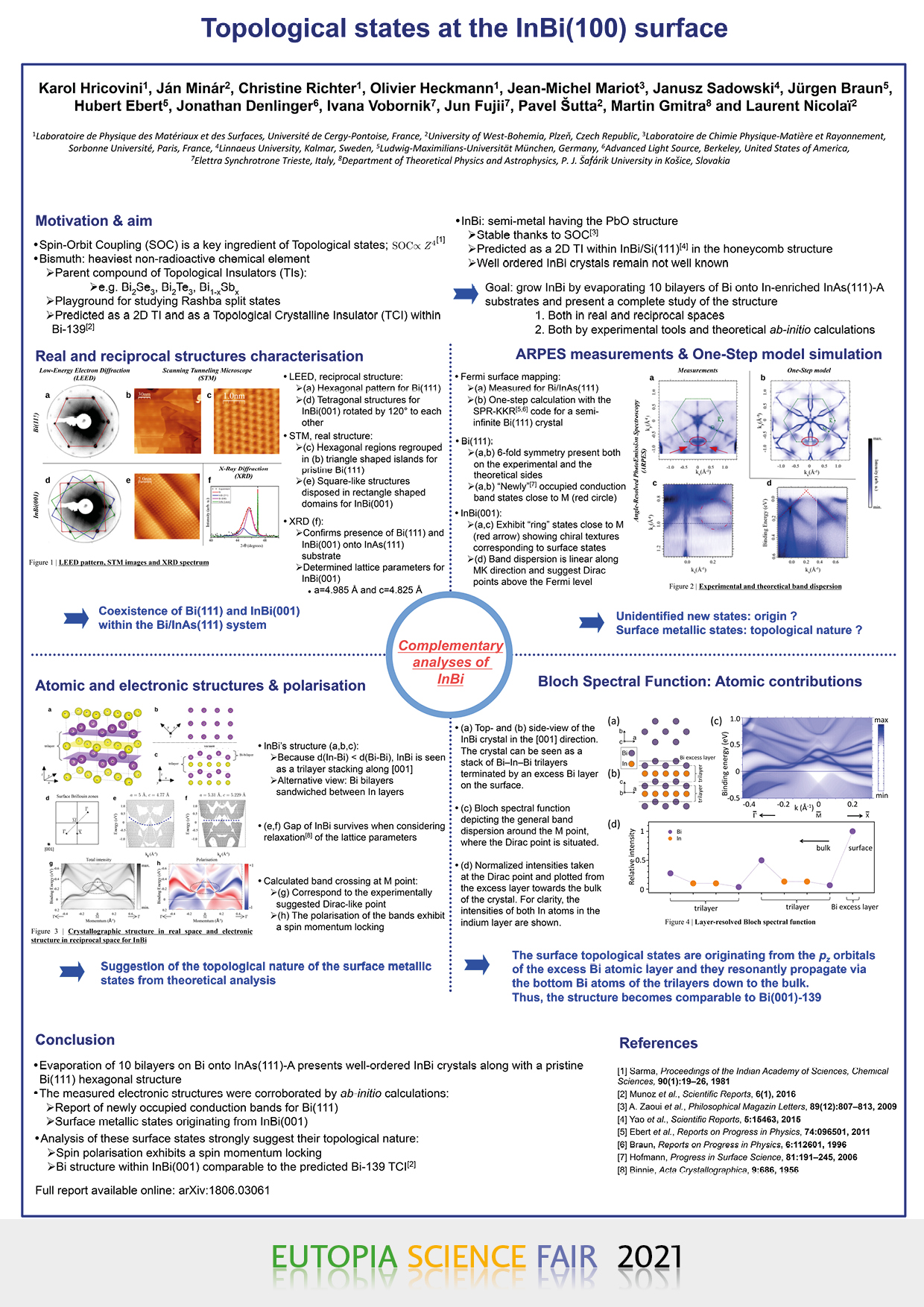
(8) Department of Theoretical Physics and Astrophysics, P. J. Šafárik University in Košice, SlovakiaThe ongoing research in topologically protected electronic states is driven not only by the obvious interest from a fundamental perspective but is also fuelled by the promising use of these non-trivial states in energy technologies such as the field of spintronics. It is therefore important to find new materials exhibiting these compelling topological features. InBi has been known for many decades as a semi-metal in which Spin-Orbit Coupling plays an important role. SOC plays a key role for emergence of novel topological states.
We present a thorough analysis of InBi, grown on InAs(111)-A surface, both, experimental by Angular-Resolved Photoemission Spectroscopy measurements and theoretical by fully-relativistic ab-initio electronic band calculations. We found existence of topologically non-trivial metallic surface states due to formed Bi bilayer with fundamental role of Bi within these electronic states. Moreover, InBi appears to be a topological crystalline insulator whose Dirac cones at the (001) surface are pinned at high-symmetry points. Consequently, as they are also protected by time-reversal symmetry, they can survive even if the in-plane mirror symmetry is broken at the surface. - De Vos et al., Vrije Universiteit Brussel
-
Development of technology for spatial three-dimensional liquid chromatography
JELLE DE VOS, THOMAS THEMELIS, ALI AMINI, SEBASTIAAN EELTINK
Vrije Universiteit Brussel, Department of Chemical Engineering, Pleinlaan 2, Brussels, Belgium
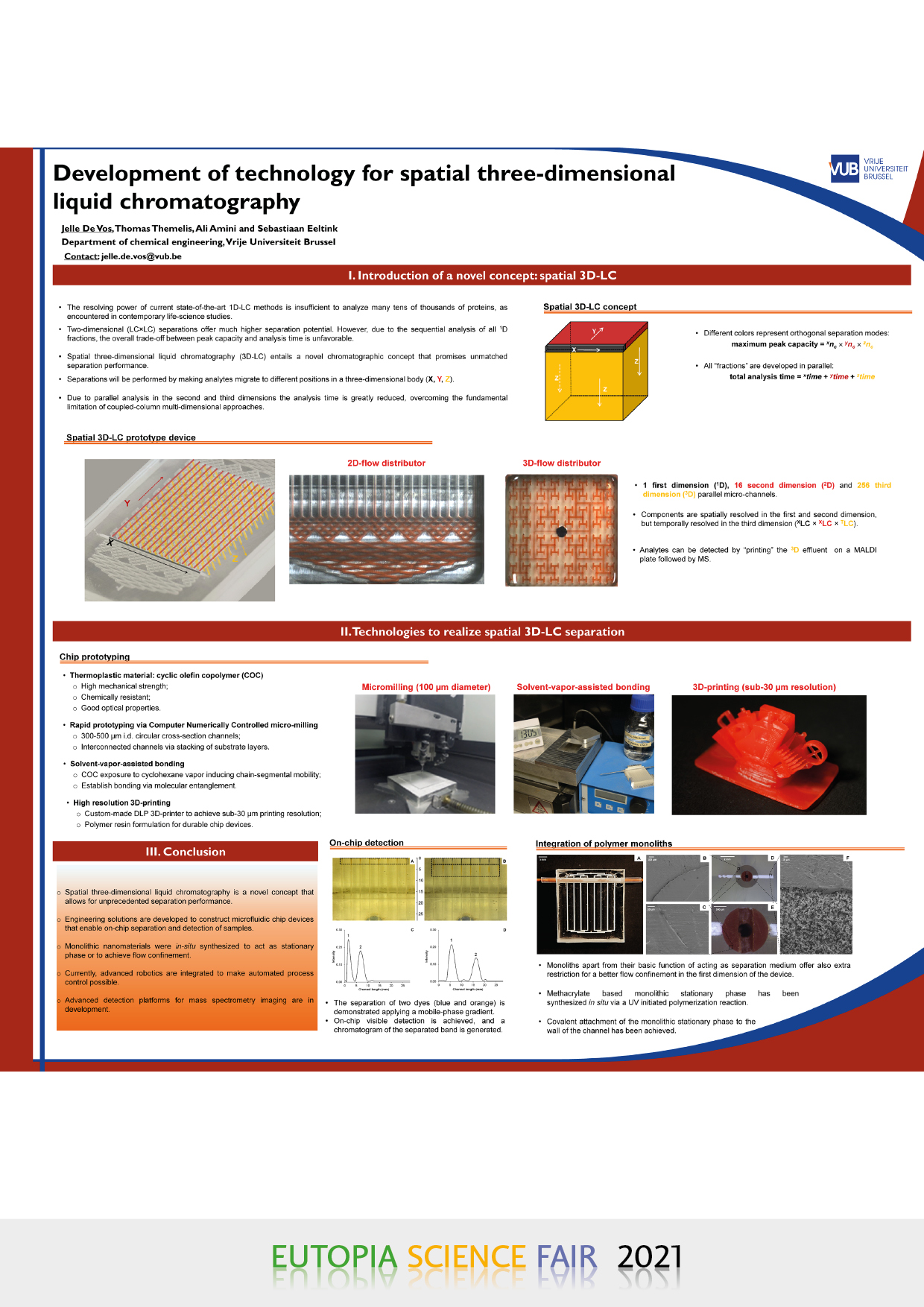
High-performance liquid chromatography (HPLC) has emerged as a dominant separation technique in the field of analytical chemistry and is widely applied in many areas, including life-science research, clinical diagnostics, and the (bio)pharmaceutical sector. For the analysis of samples used in biomarker discovery studies, which are typically characterized by a large sample complexity (often containing over 1,000,000 compounds) and broad dynamic range, the current state-of-the-art HPLC technology does not allow to fully resolve the analytes. Column-based multidimensional LC approaches, i.e., by coupling columns with complementary (orthogonal) separation mechanisms, have been developed to improve the separation performance. In this separation platform, where the fractions originating from multiple columns are sequentially analyzed, a single analysis takes a long time to complete. This makes the technology intrinsically unsuitable for high-throughput screening.
To advance the separation performance, and to overcome this bottleneck of sequential runs, the development of a microfluidic separation device for spatial 3D-LC was explored. In spatial 3D-LC chromatography components are separated inside the microchannels of the device with each peak being characterized by its X, Y, and Z coordinates in the separation body. We investigated different design aspects of a microfluidic device for comprehensive spatial 3D-LC, including flow distributor design and channel layout. During the different developments in X-, Y-, and Z-direction, the analytes and therefore the flow should not convolute in other dimensions. To this end, we developed active flow confinement technology, which also allows to modulate the mobile-phase composition between the 1st and 2nd dimension development. To allow for automated process operation, mechanisms applying advanced robotics have been successfully integrated on-chip. Furthermore, approaches to integrate functionalized monolithic stationary phases at pre-defined locations in microfluidic device have been realized, which are essential to realizing a spatial 3D-LC separation of complex samples.
- O. Fichet, CY Cergy Paris Université
-
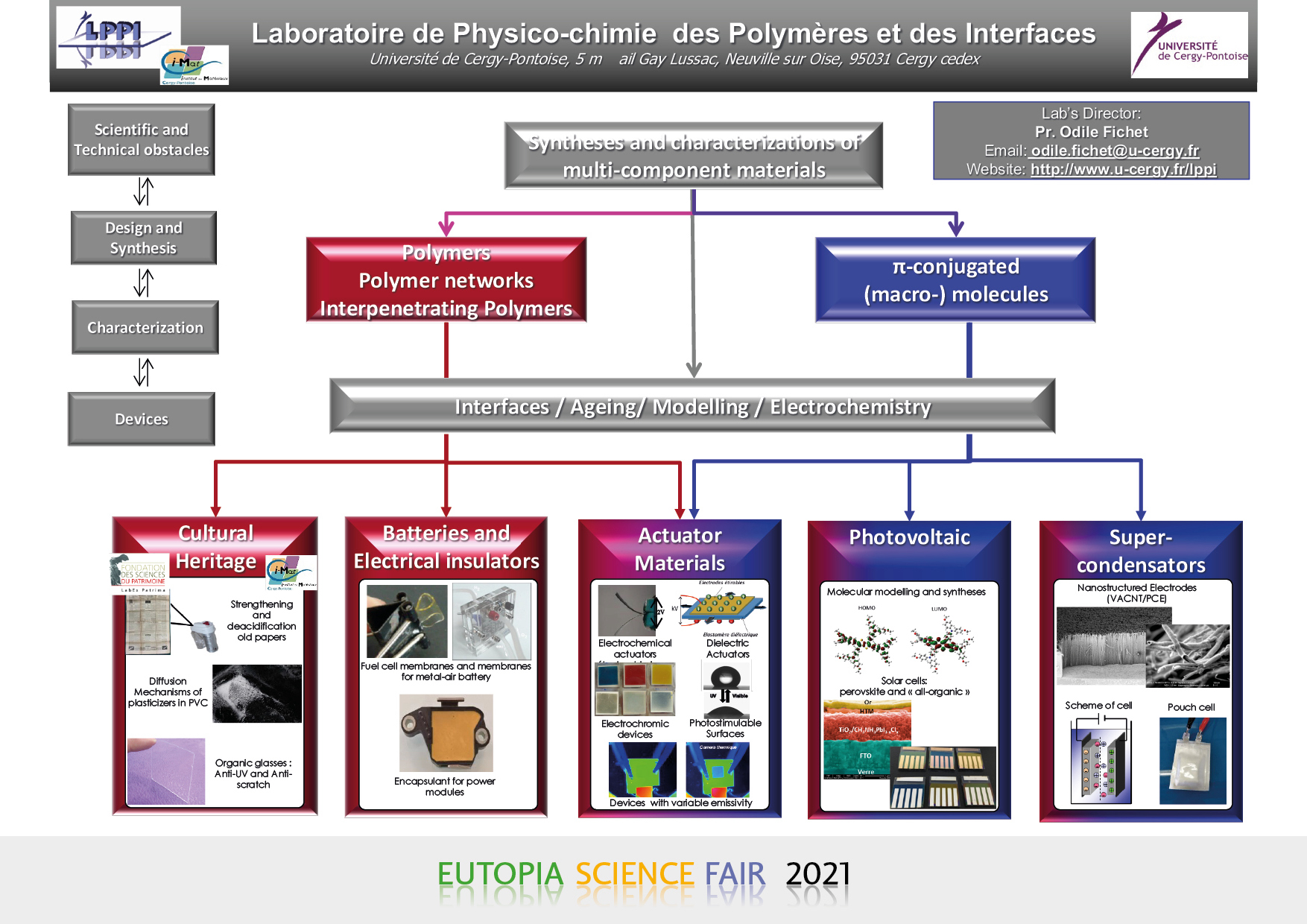
Laboratoire de Physico-chimie des Polymères et des Interfaces
- Gwendoline Petroffe et al., CY Cergy Paris Université
-
Adaptive Thermal Control of Satellites by ElectroActive Polymeric Coating
- C. Plesse et al., CY Cergy Paris Université
-
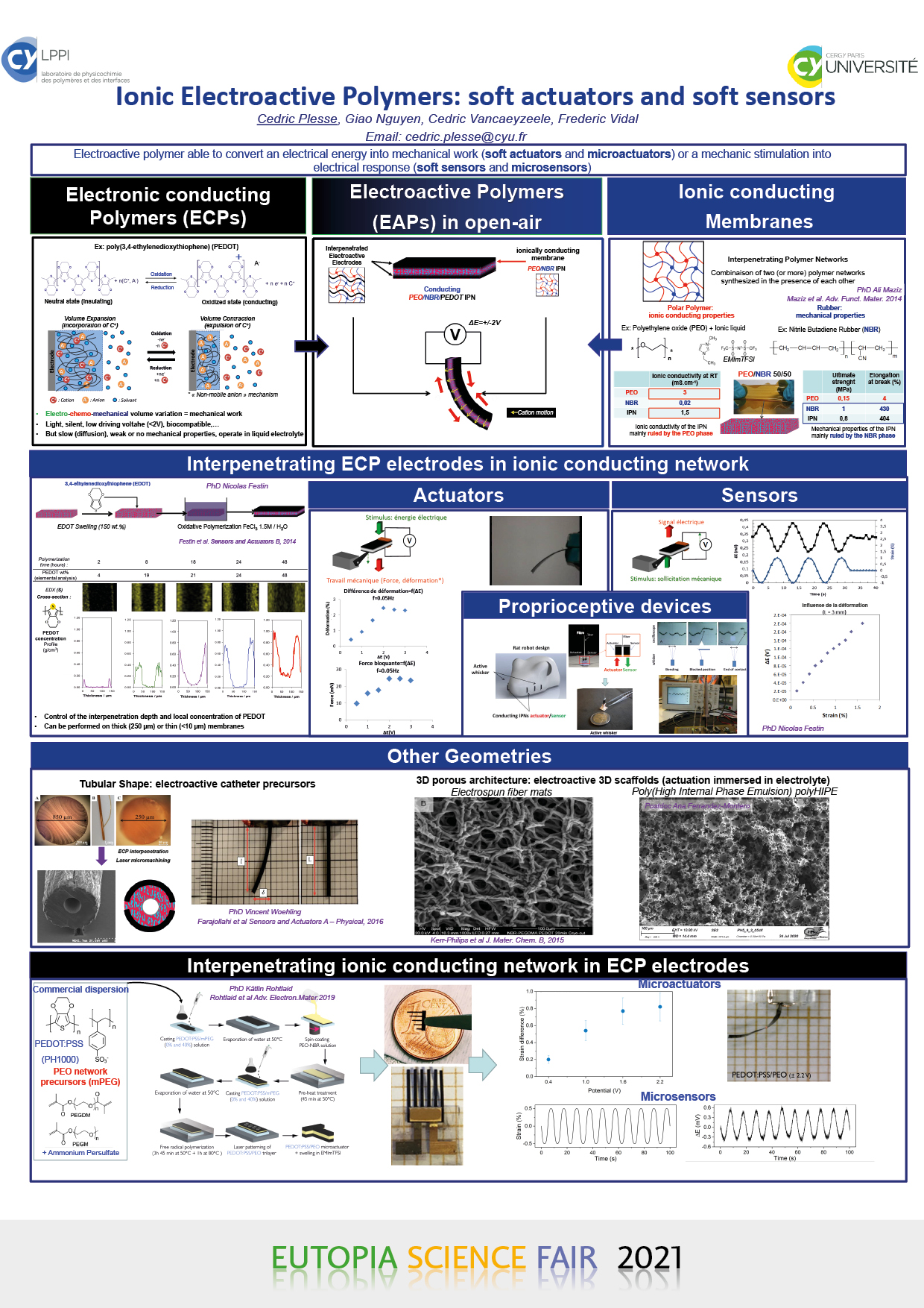
Ionic Electroactive Polymers : soft actuators and soft sensors
CEDRIC PLESSE , GIAO NGUYEN , CEDRIC VANCAEYZEELE , FREDERIC VIDAL
CY Cergy Paris Université, LPPI, Chemistry dpt, 95000 CERGY, FRANCE
Ionic ElectroActive Polymers (EAPs) are smart materials which respond to an electrical stimulus by undergoing volume variation due to ion motions. Since the first works on these materials in the early 90’s, numerous concepts have been described and have stimulated the imagination of chemists, physicists, biologists and mechanical engineers since they make possible the development of biomimetic soft actuators and soft sensors, precursors of so-called artificial muscles, with numerous potential applications in soft robotics, microelectromechanical systems, biomedical devices or smart textiles. Electronic conducting polymers (ECPs) belongs to the family of ionic EAPs and present numerous advantages such as being lightweight, soft, biocompatible and activated by low voltage (<2V). Their actuation principle relies on the ECP volume variation induced by ion exclusion/expulsion when submitted to a redox process in the presence of an electrolyte. Combining ECP electrodes and ionic conducting membranes allows developing trilayer devices able to operate in open-air and to (i) convert electrochemical stimulation into mechanical work (actuator) or (ii) to detect and quantify mechanical stimulation through output electrical signal (sensor).
The performances of such devices are mainly determined by the interdependent properties of the two partners: ECP electrodes (electronic conductivity and electroactivty) and ionic conducting membranes (ionic conductivity and mechanical robustness). We described here the work developed in LPPI (CY) on versatile electroactive devices, able to contract, expand, bend or sense by the synthesis of conducting Interpenetrating Polymer Network (IPN) architecture allowing to combine and tune these properties. According to this strategy, it has been possible to elaborate efficient electroactive materials but also to miniaturize them with classical microsystem processes allowing the development of soft (micro-)actuators and sensors. Finally, elaborating these materials with various shapes allowed the development of innovative biomedical applications such as electroactive catheter precursors with electrocontrollable bending or 3D porous electroactive scaffolds (electrospun fiber mats or polyHIPEs) with electrotunable porosity for cell mechanotransduction and tissue engineering.
- M. Vigroux et al., CY Cergy Paris Université
-
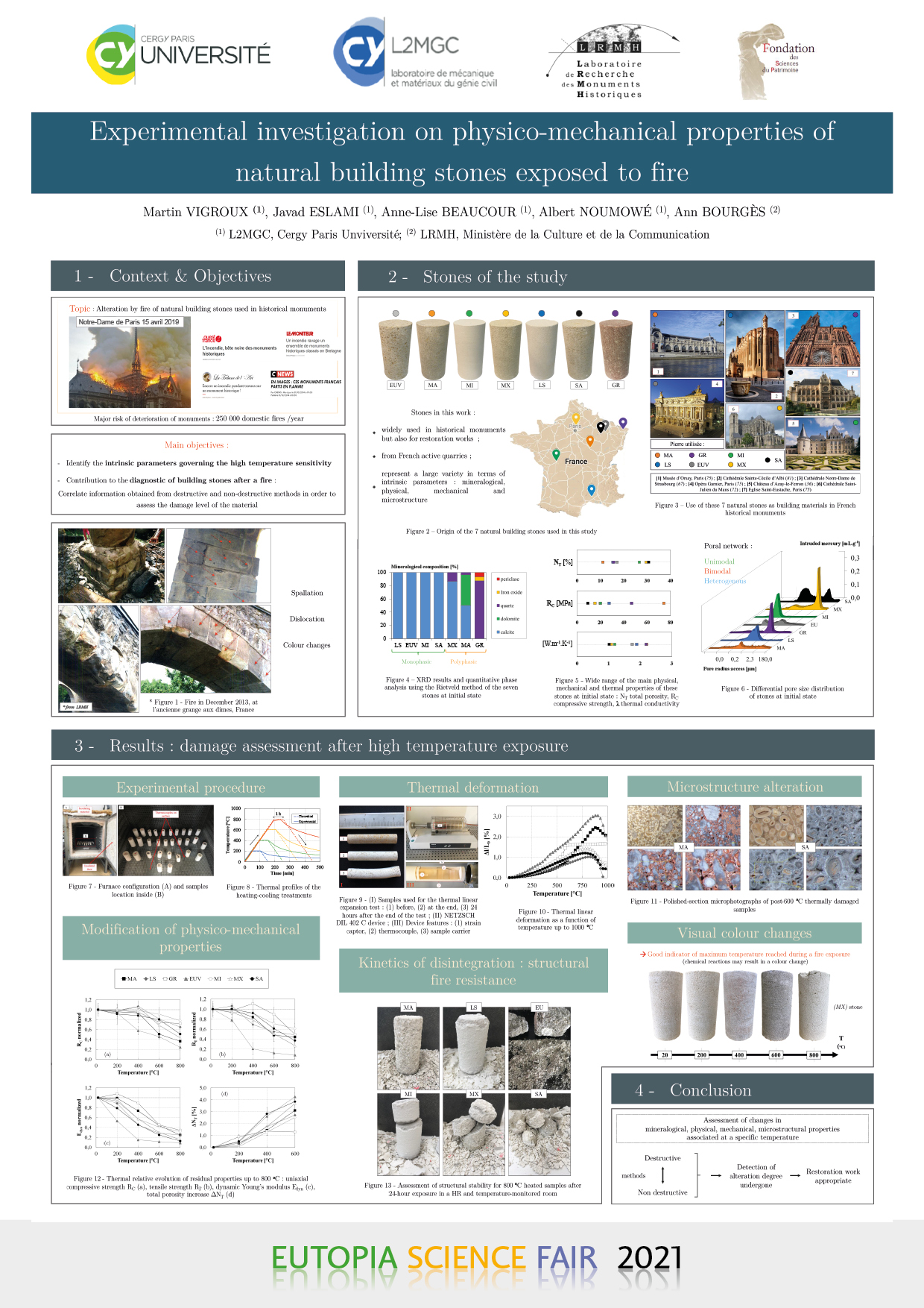
Experimental investigation on physico-mechanical properties of natural building stones exposed to fire
MARTIN VIGROUX (1), JAVAD ESLAMI (1), ANNE-LISE BEAUCOUR (1), ALBERT NOUMOWÉ (1) ANNE BOURGÈS (2)
(1) L2MGC, CY Cergy Paris Université
(2) LRMH, Ministère de la Culture et de la Communication, FranceFire appears as one of the main causes of built heritage weathering because it can generate irreversible damage with long-lasting effects, in a very short period of time. A well-known example for that is the recent fire that devastated Paris‘ world-famous Notre–Dame Cathedral on April 15th 2019.
This study focuses on the the study of the high temperature behaviour of various natural stones, used as building materials in the built heritage. Firstly, experimental measurements under high temperature allowed the identification of physico-chemical phenomena occurring during heating and cooling. Thus, the influence of mineralogy on the thermo-chemical stability of limestones and sandstone has been discussed. In addition, thermal deformation measurements up to 1050 °C have highlighted the role of certain petrophysical parameters on the mechanical behaviour of these stones at high temperatures. The evolution of thermal properties during heating has been determined. In addition, the evolution of residual properties (compressive strength, tensile strength, dynamic modulus of elasticity, total porosity, water capillary coefficient) of these stones after heating-cooling cycles up to 800 °C was experimentally determined. Microscopic observations, coupled with mercury porosimetry analyses, allowed the assessment of the porous network modification. The results of this study, obtained using different experimental methods, contribute to the diagnosis of stone-heritage structures that have suffered a fire. On the one hand, the consequences of the post-fire structural capacity have been established and, on the other hand, the consequences on its durability facing various environmental attacks have been analyzed. - S.Dahle, University of Ljubljana
-
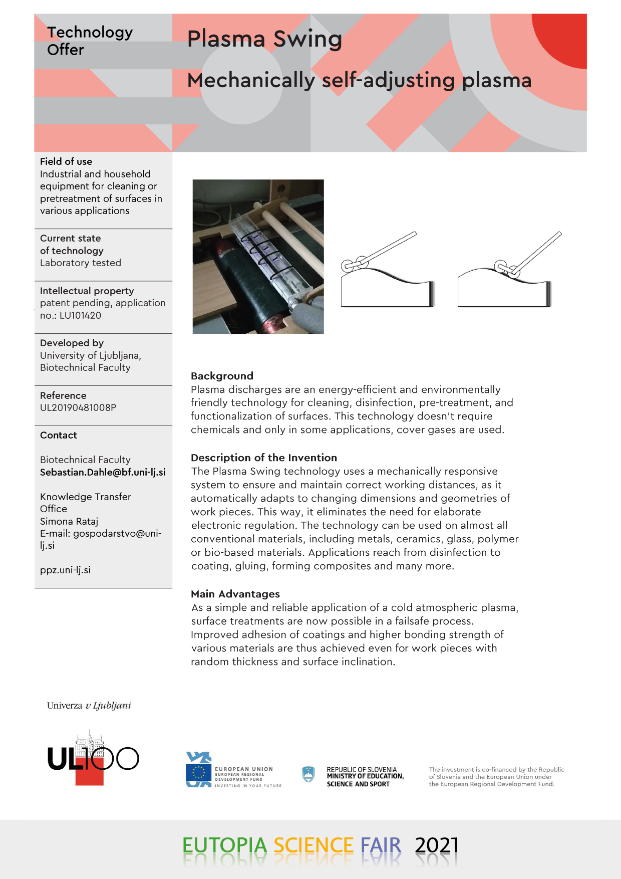
Plasma Swing, Mechanichally self-adjusting plasma
- 2. The World of Life: Chemistry, Genetics, Medicine. Part 1
-
- C. Zanato et al., CY Cergy Paris Université
-
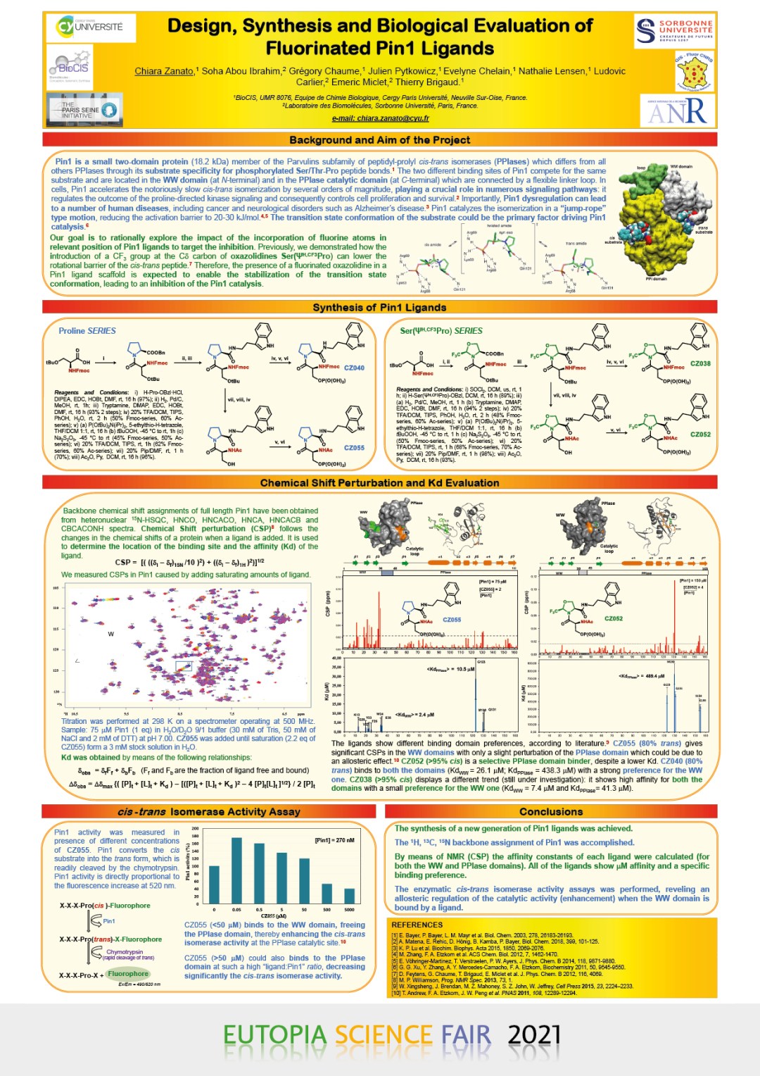
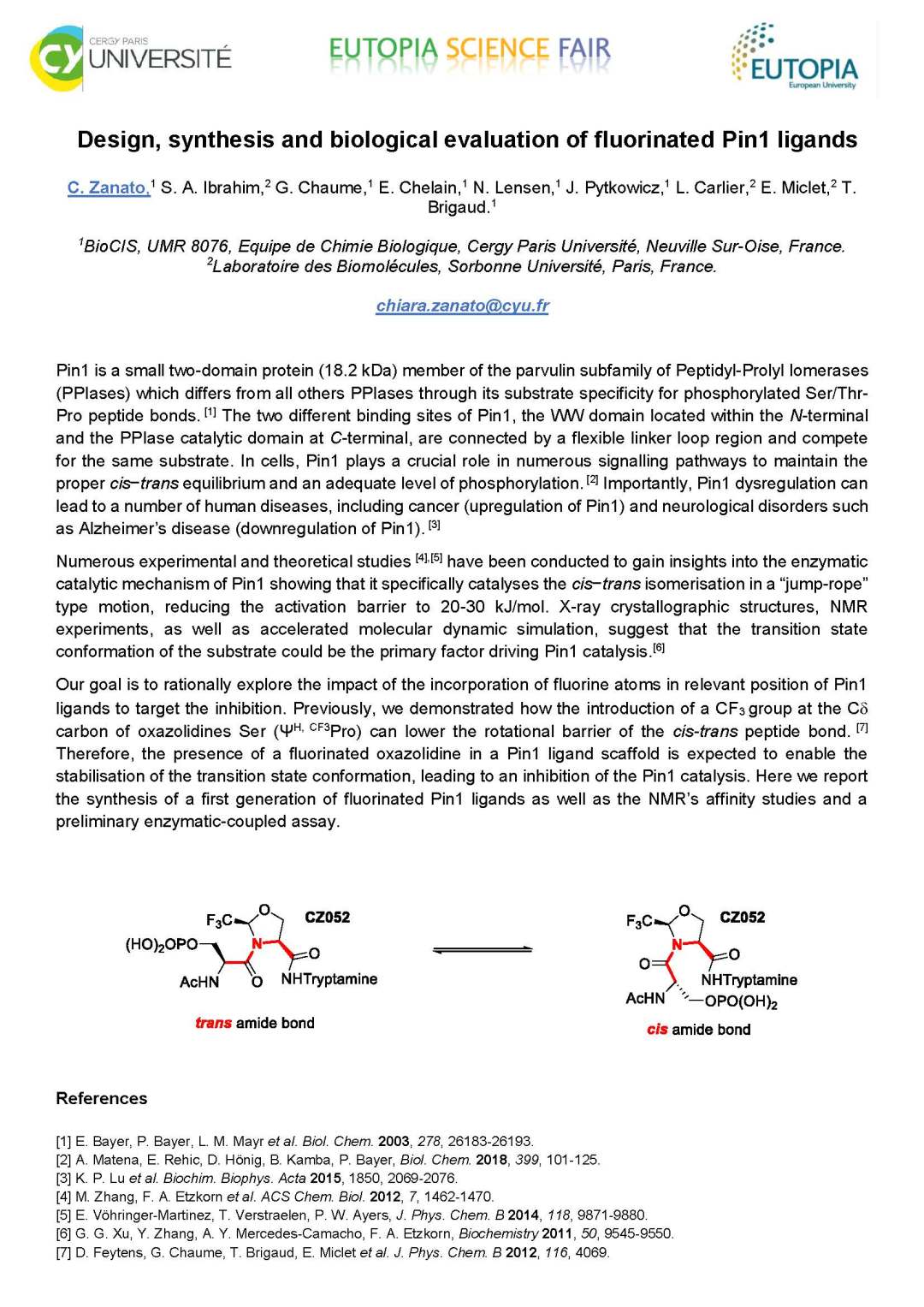
Design, synthesis and biological evaluation of fluorinated Pin1 ligands
- K. Wood, University of Warwick
-
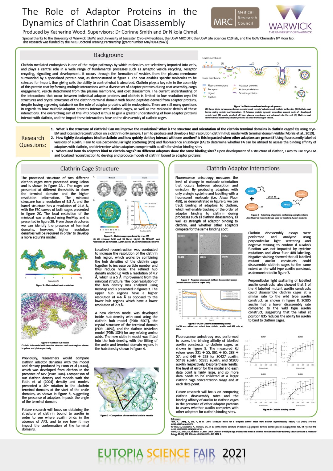
Development of a Cryo-EM Clathrin Structure for Assessing Clathrin Adaptor Interactions
KATHERINE WOOD
University of WarwickClathrin-mediated endocytosis is one of the major pathways by which molecules are selectively imported into cells and plays a central role in a wide range of fundamental processes such as synaptic vesicle recycling, receptor recycling, signalling and development. It occurs through the formation of vesicles from the plasma membrane surrounded by a specialized protein coat. The coat enables specific molecules to be selected for import, thus giving cells the ability to control what is absorbed. Clathrin plays a key role in the assembly of this protein coat, forming multiple interactions with a diverse set of adaptor proteins as the coat assembles, engages with the cargo destined for endocytosis, facilitates detachment of the vesicle from the membrane and finally disassembles from the newly detached vesicle.
This research project aims to assess the interactions of adaptor proteins with clathrin, and how they impact clathrin coat disassembly. Despite the publication of several cryo-EM clathrin cage structures with bound adaptors, no clathrin cage control has been published so far. Development of a control is important for validating the presence of adaptor densities bound to clathrin, as well as conformational changes of clathrin that may occur with the adaptors bound. We have produced cryo-EM maps of two types of clathrin cage and reconstructed the hub regions to generate a higher resolution model. Our next goal is to develop a higher resolution structure of clathrin bound to the disassembly adaptor protein auxilin to determine where auxilin binds. We are also using fluorescence anisotropy and stopped flow experiments to analyse the binding kinetics of different adaptors to clathrin cages, in order to understand their affinity for clathrin and whether they compete for binding.
- H. G. Smith, University of Warwick
-
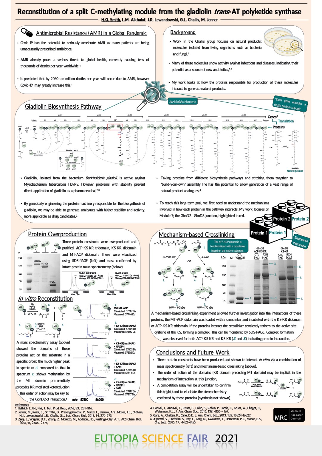
Reconstitution of a Split C-Methylating Module from the Gladiolin trans-AT Polyketide Synthase
HELEN G. SMITH(1,2), LONA ALKHALAF(2), JÓZEF R. LEWANDOWSKI(2), GREGORY L. CHALLIS(2,3,4), MATTHEW JENNER(2,4)
(1) Warwick Medical School, University of Warwick, Coventry, UK,
(2) Department of Chemistry, University of Warwick, Coventry, UK,
(3) Department of Biochemistry and Molecular Biology, Monash University, Clayton, Australia,
(4) Warwick Integrative Synthetic Biology Centre, University of Warwick, Coventry, UKPolyketides are a class of natural product which show great potential as a source of new antibiotics. They are active against several infectious diseases and many are already in use as pharmaceuticals. Despite the great potentialof polyketides several problems often prevent their direct application as pharmaceuticals, including stability, bioavailability, toxicity and resistance. It is possible to overcome these issues through structural modification of the molecules. This can either be achieved through chemical synthesis, which is made difficult by the complex nature of the molecules, or through genetic engineering of the protein machinery responsible for the biosynthesis of thecompounds.
Modular polyketide synthases (PKSs) biosynthesise many medicinally active polyketides. Each PKS moduleis composed of several independently folded catalytic domains that incorporate a building block into the polyketidechain and modify the α- and β-carbons of the resulting acyl carrier protein (ACP)-bound thioester. Modular PKSs typically consist of several protein subunits, which interact via structurally defined docking domains (DDs) at their N- and C-termini. DDs have been exploited in the genetic engineering of PKSs to produce novel natural product analogues; for examples in the DEBS PKS which synthesises the polyketide core of erythromycin A.
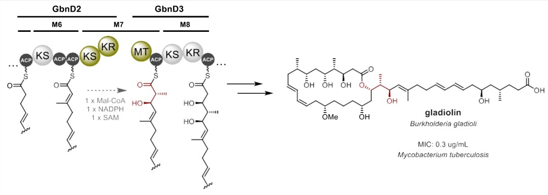
Recent work identified gladiolin (Figure 1), a polyketide with activity against Mycobacterium tuberculosis (MIC: 0.4 μg / mL) from Burkholderia gladioli BCC0238. Bioinformatics analyses identified the intersubunit junction within module 7 of the gladiolin PKS (figure 1) as lacking docking domains. This is the first identified example of a trans-AT PKS interface lacking DDs. Here we report work directed towards dissecting and understanding this novel intersubunit interaction interface, which will ultimately aid biosynthetic engineering experiments to produce novel analogues of gladiolin.
Figure 1. Partial biosynthetic scheme for the production of gladiolin. The KR/MT interface separating the GbnD2 and GbnD3 subunits is highlighted in green and the chemical transformations across this boundary are highlighted in red on the biosynthetic intermediate and the structure of gladiolin.
- D. Miklavčič, University of Ljubljana
-
Electroporation in research, biotechnology and clinical use
- L. Peterlin Masic, University of Ljubljana
-
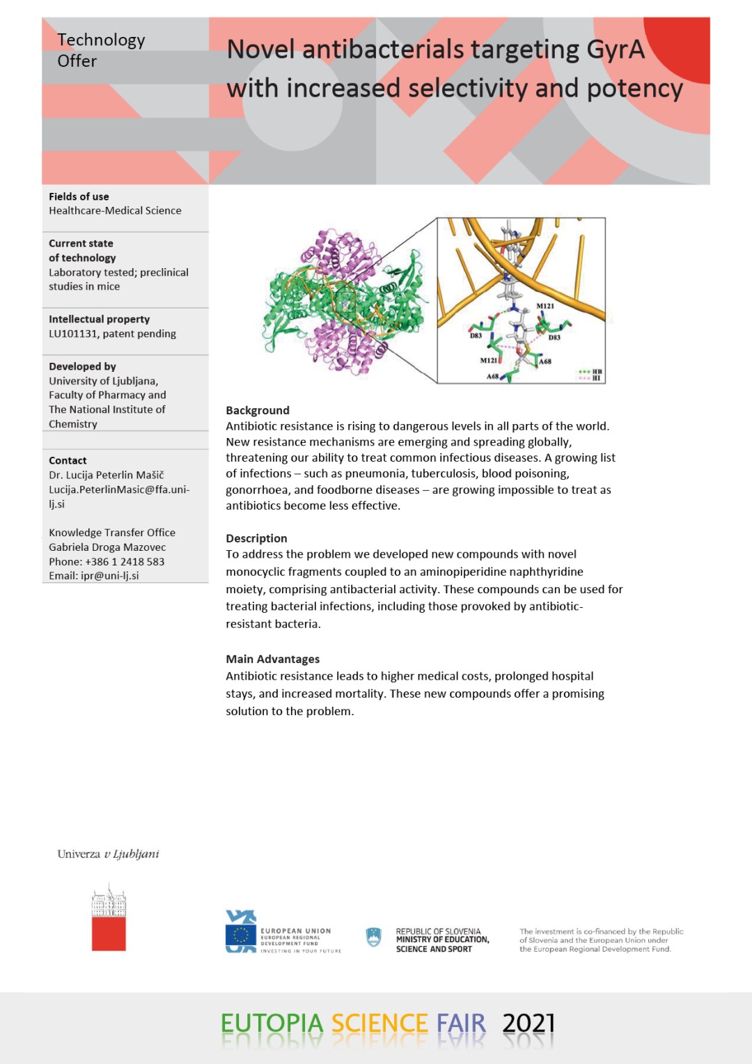
Novel antibacterials targeting GyrA with increased selectivity and potency
- Yean Ming C. et al., University of Warwick
-
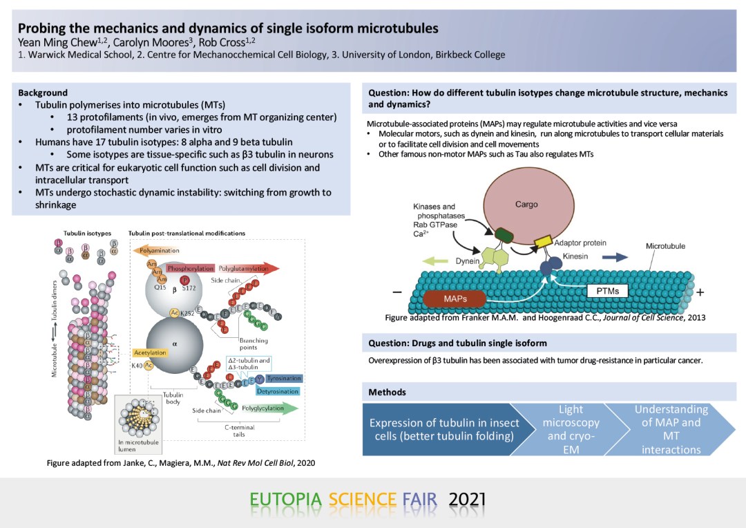
Probing the mechanics and dynamics of single isoform microtubules
YEAN MING CHEW (1,2), CAROLYN MOORES(3), ROB CROSS(1,2)(1) Warwick Medical School
(2) Centre for Mechanochemical Cell Biology
(3) University of London, Birkbeck CollegeMicrotubules are hollow cylindrical structures in cells which participate in cell motility, cell division and intracellular transport. This structure is made of 13 protofilaments in vivo, of which the individual protofilament is a polymer heterodimers of alpha and beta tubulin, joined in a head-to-tail fashion. Microtubules have characteristic property – switching from growth phase to shrinking phase stochastically and vice versa which involves the addition and loss of tubulin heterodimers respectively.
In human, there are eight alpha- and nine beta-tubulin isotypes which share high similarity in protein sequence. Some are tissue-specific, such as b3 tubulin, which is highly expressed in some cancer cells, has been found to be less sensitive to cancer drug, taxol, in particular cancer.
Microtubules and microtubule-associated proteins (MAPs) regulate each other. Our previous finding revealed that kinesin-1, a molecular motor which is responsible for intracellular transport, expands and stabilises microtubule lattice. Together with molecular motors, microtubules serve as train track to allow intracellular transport and some other cell activities.
Currently, most studies are carried out using tubulin extracted from bovine or porcine brains, which are composed of a mixture of tubulin isotypes. Therefore, there is a need to study individual tubulin isotypes to gain understanding of how these small differences in protein sequence affect cells.
We are investigating whether lattice expansion and stabilisation of microtubules conferred by kinesin-1 can be affected by different tubulin isotypes. By overexpressing his-tagged and FLAG-tagged a– and b– tubulin respectively, in insect cells, single-isotype recombinant tubulin can be obtained. The dynamics of microtubules made of single-isotype tubulin in the presence of kinesin can also be observed under dark field microscope without the use of fluorescently-labelled tubulin which could potentially affect microtubule dynamics.
- J. Furner-Pardoe et al., University of Warwick
-
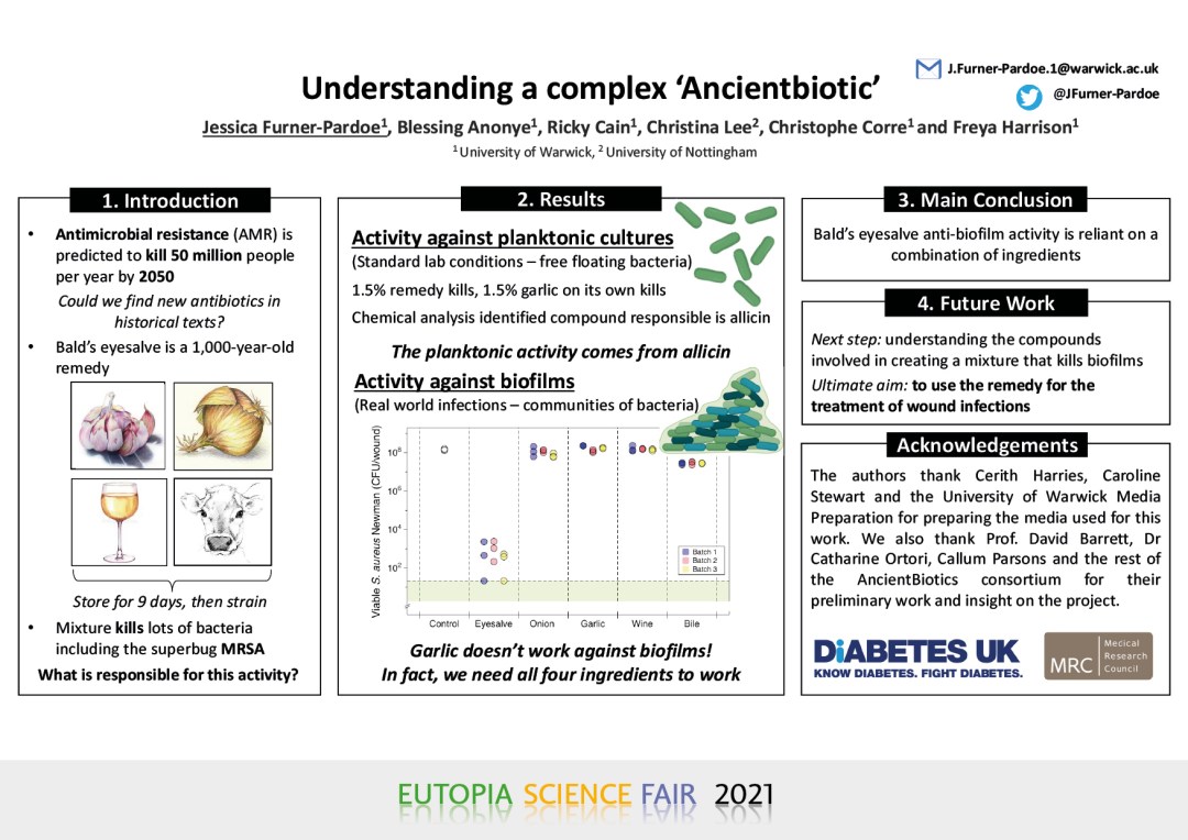
Anti-biofilm activity of 1,000-year-old-remedy requires the combination of multiple ingredients
JESSICA FURNER-PARDOE (1), BLESSING O ANONYE (1‡),, RICKY CAIN (1,#), JOHN MOAT (1), CATHERINE A. ORTORI (2), CHRISTINA LEE (2), DAVID A. BARRETT (2), CHRISTOPHE CORRE1 AND FREYA HARRISON (1)
(1) University of Warwick, UK.
(2) University of Nottingham, UK. ‡Present address: University of Central Lancashire, UK.
(#) Present address: Evotec (U.K.) Ltd., Oxfordshire, UK.
Combatting the rise in antibiotic resistance is one of the major challenges in modern science. Studying historical medical remedies could help reveal new antibiotics. Historical medical manuscripts prescribe complex preparations of several ingredients to treat infections, and it is suspected their efficacy may rely on creating a cocktail of natural products. A reconstructed 1000-year-old remedy, containing onion, garlic, wine, and bile salts, was previously shown to kill methicillin-resistant Staphylococcus aureus in a mouse chronic wound model, and Pseudomonas aeruginosa in an ex vivo chronic lung infection model. Recently, we have shown this activity extends to biofilms of various ESKAPE pathogens, including MRSA and Acinetobacter baumannii. Traditionally, natural product research has focussed on isolating active compounds using planktonic cultures, however, here we present data that by doing so we may overlook efficacious mixtures. The potent activity against planktonic cultures could be achieved with garlic alone, and chemical analysis has identified the compound responsible to be allicin. However, the garlic alone has no activity against biofilms of the same bacteria. In fact, all four ingredients were necessary to get full potent antibiofilm activity. This highlights the importance of interactions within plant mixtures. By incorporating biofilm studies earlier in the search of active compounds, we may highlight potent interactions and generate promising mixtures for the treatment of infections.
- R. García Maset, University of Warwick
-
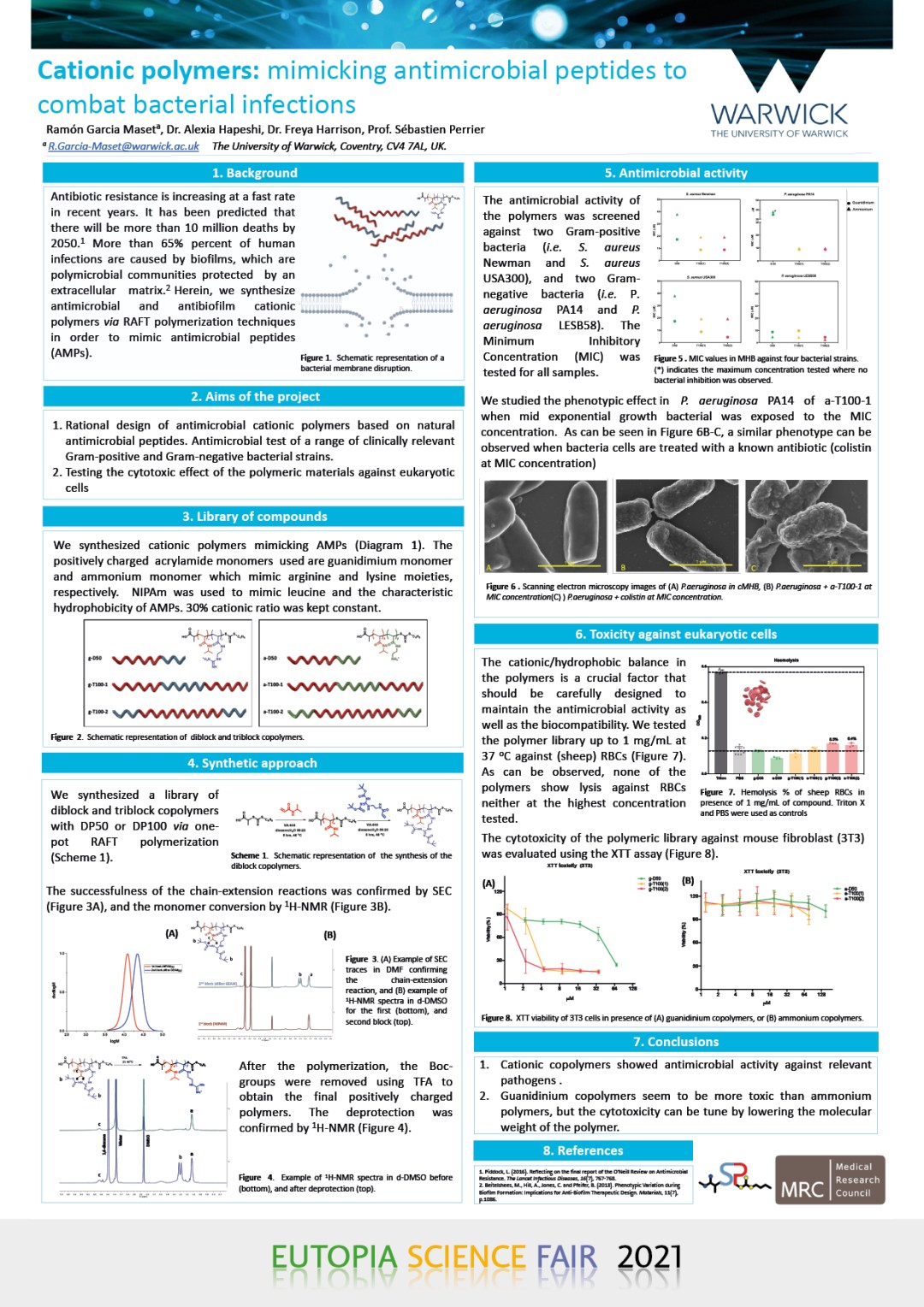
Cationic polymers show antimicrobial and antibiofilm activity against multidrug resistant S. aureus
RAMON GARCIA MASET
Warwick Medical School MRCDTP
The rational design of new antibiotic drugs is of imminent global necessity: by 2050 more than 10 million deaths and an immense cost for the worldwide health systems are predicted due to multidrug bacterial infections. Recently, antimicrobial peptides (AMPs) have shown promising antimicrobial activity against a broad range of pathogens. However, only a few have reached clinical trials due their cytotoxicity and highly cost production.Herein, we aimed to mimic and improve the limitations of AMPs by using polymer chemistry. We designed a family of cationic polymers synthetized by reversible addition-fragmentation chain transfer (RAFT) polymerization. The family of polymers differs from each other in their molecular weight and the segregation of the cationic content along the backbone. They showed good antimicrobial activity against multidrug-resistant S. aureus strains in standard culture conditions and using culture conditions similar to those found in wound infections. None of the synthetized polymers showed hemolysis against red blood cells. Additionally, low molecular weight polymers did not show toxicity against fibroblast cells. The activity of one of the polymers was tested against S. aureus biofilms in a collagen wound model. A complete biofilm inhibition was observed when the artificial wound was pre-treated with the polymer. Cationic polymers offer the possibility to mimic an existing natural product in order to establish a new category of antibiotics with a future scope in clinics.
- S. Li et al., University of Warwick
-
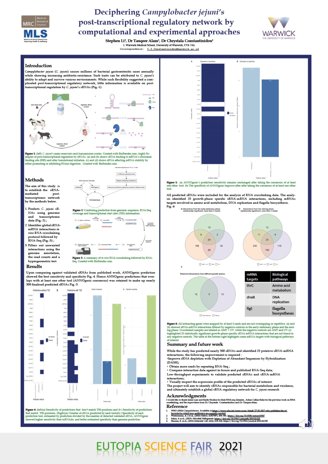
Decyphering Campylobacter jejuni’s post-transcriptional regulatory network by computational and experimental approaches
STEPHEN LI1, DR TAUQEER ALAM1, DR CHRYSTALA CONSTANTINIDOU1
1 Warwick Medical School, University of Warwick, CV4 7AL C.I.Constantinidou@warwick.ac.ukCampylobacter jejuni (C. jejuni) causes millions of bacterial gastroenteritis cases each year worldwide, while also showing increasing antibiotic- resistance. Given its medical and public health importance, it is remarkable that C. jejuni is one of the least understood enteropathogens. In particular, very little information is available on post-transcriptional regulation by small non-coding RNAs (sRNAs), despite being directly associated with virulence and antibiotic resistance in other pathogens.
Previous studies have only experimentally validated about 20 C. jejuni sRNAs, whilst their binding targets remain elusive. Hence my project aims to predict unannotated small RNAs in C. jejuni genome using machine learning tools and RNA-Seq coverage analysis. The predicted sRNAs facilitates the subsequent analysis of in-house RNA-Seq data from over 20 infection and transmission relevant conditions and my own in vivo RNA crosslinking experiment.
The project ultimately aims to identify and reconstruct condition-specific sRNA-mRNA interaction networks. The result will provide the C. jejuni research community with a better understanding of how C. jejuni regulates growth, survival and virulence. This result will also expand sRNA research into a broader range of pathogen species.
- C. J. Holmberg et al., University of Gothenburg
-
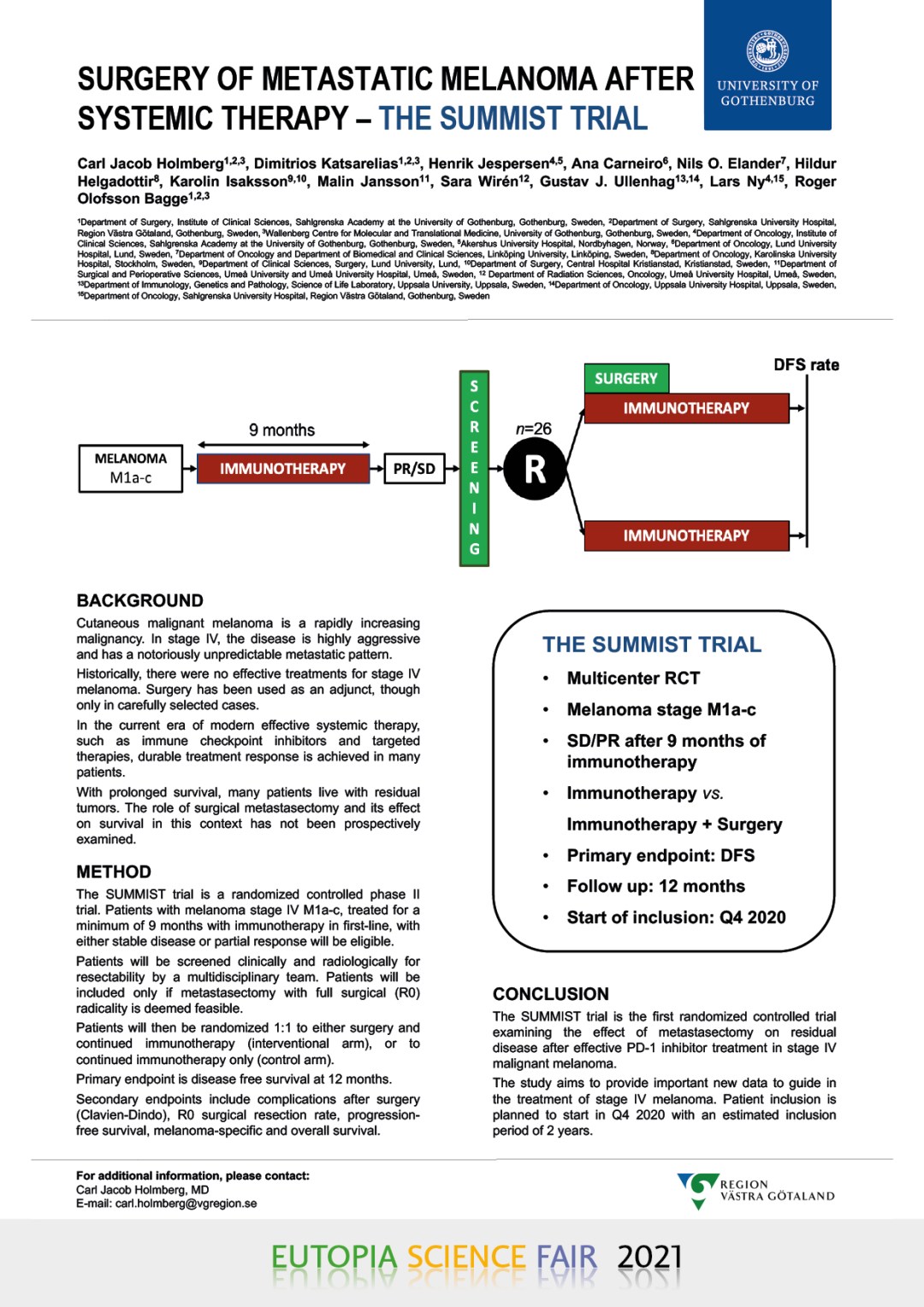
Surgery of metastatic melanoma after systemic therapy – the SUMMIST trial: study protocol for a randomized controlled trial
CARL JACOB HOLMBERG (1,2,3), DIMITRIOS KATSARELIAS (1,2,3), HENRIK JESPERSEN (4,5), ANA CARNEIRO (6), NILS O. ELANDER (7), HILDUR HELGADOTTIR (8), KAROLIN ISAKSSON (9,10), MALIN JANSSON (11), SARA WIRÉN (12), GUSTAV J. ULLENHAG (13,14), LARS NY (4,5), ROGER OLOFSSON BAGGE (1,2,3)
(1) Department of Surgery, Institute of Clinical Sciences, Sahlgrenska Academy at the University of Gothenburg, Gothenburg, Sweden.
(2) Department of Surgery, Sahlgrenska University Hospital, Region Västra Götaland, Gothenburg, Sweden.
(3) Wallenberg Centre for Molecular and Translational Medicine, University of Gothenburg, Gothenburg, Sweden.
(4) Department of Oncology, Institute of Clinical Sciences, Sahlgrenska Academy at the University of Gothenburg, Gothenburg, Sweden.
(5) Department of Oncology, Sahlgrenska University Hospital, Region Västra Götaland, Gothenburg, Sweden
(6) Department of Oncology, Lund University Hospital, Lund, Sweden.
(7) Department of Oncology and Department of Biomedical and Clinical Sciences, Linköping University, Linköping, Sweden.
(8) Department of Oncology, Karolinska University Hospital, Stockholm, Sweden
(9) Department of Clinical Sciences, Surgery, Lund University, Lund.
(10) Department of Surgery, Central Hospital Kristianstad, Kristianstad, Sweden
(11) Department of Surgical and Perioperative Sciences, Umeå University and Umeå University Hospital, Umeå, Sweden.
(12) Department of Radiation Sciences, Oncology, Umeå University Hospital, Umeå, Sweden.
(13) Department of Immunology, Genetics and Pathology, Science of Life Laboratory, Uppsala University, Uppsala, Sweden.
(14) Department of Oncology, Uppsala University Hospital, Uppsala, Sweden.
Correspondence to: Carl Jacob Holmberg, MD – ORCID iD: https://orcid.org/0000-0002-3512-4535Department of Surgery, Institute of Clinical Sciences, Sahlgrenska Academy at the University of Gothenburg, Sahlgrenska University Hospital, Gothenburg, Sweden.E-mail: carl.holmberg@vgregion.se
Introduction: Cutaneous malignant melanoma is a rapidly increasing malignancy. In stage IV, the disease is highly aggressive and has a notoriously unpredictable metastatic pattern. Historically, there were no effective treatments for stage IV melanoma. Surgery has been used as an adjunct, though only in carefully selected cases. In the current era of modern effective systemic therapy, such as immune checkpoint inhibitors and targeted therapies, durable treatment response is achieved in many patients. With prolonged survival, many patients live with residual tumors. The role of surgical metastasectomy and its effect on survival in this context has not been prospectively examined.
Material and methods: The SUMMIST trial is a randomized controlled phase II trial. Patients with melanoma stage IV M1a-c, treated for a minimum of 9 months with immunotherapy in first-line, with either stable disease or partial response will be eligible. They will be screened clinically and radiologically for resectability by a multidisciplinary team. Patients will be included only if metastasectomy with complete removal of residual disease and full surgical (R0) radicality is deemed feasible. Patients will then be randomized 1:1 to either surgery and continued immunotherapy (interventional arm), or to continued immunotherapy only (control arm). Primary endpoint is disease free survival at 12 months. Secondary endpoints are serious adverse events, complications after surgery according to Clavien-Dindo classification and Comprehensive Complications Index, R0 surgical resection rate according to pathology report, progression-free survival, melanoma-specific survival and overall survival.
Discussion: The SUMMIST trial is the first randomized controlled trial examining the effect of metastasectomy on residual disease following effective immunotherapy in stage IV malignant melanoma. The trial is a small but important step in expanding treatment options in this patient group. Patient inclusion started Q4 2020.
KEYWORDS: MELANOMA, SURGERY, METASTASECTOMY, METASTATIC SURGERY, IMMUNOTHERAPY, PD1-INHIBITOR
- P. Muñoz-Cánoves, University of Pompeu Fabra
-
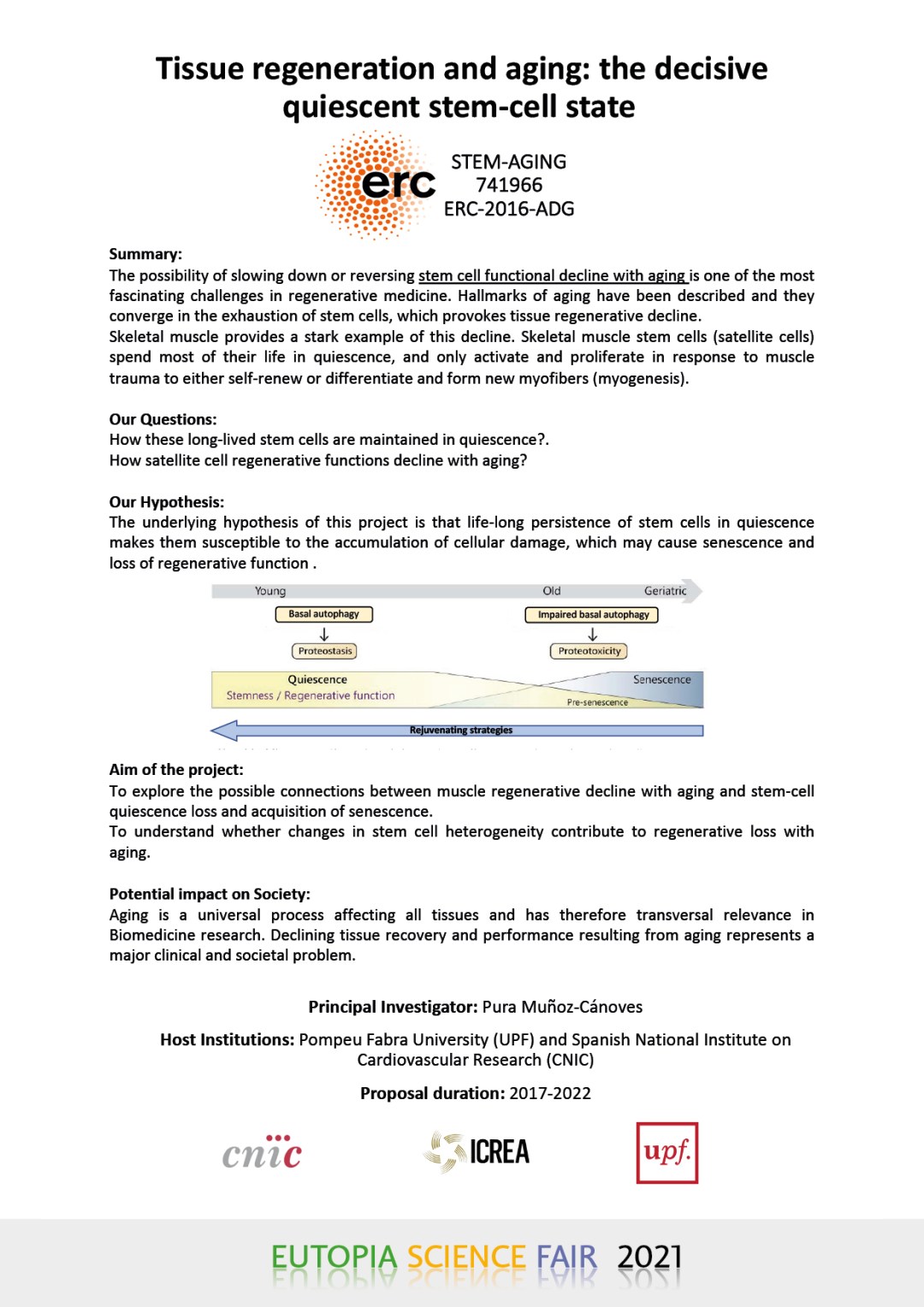
Tissue regeneration and aging: the decisive quiescent stem-cell state STEM-AGING 741966 ERC-2016-ADG
PURA MUÑOZ-CÁNOVES
Pompeu Fabra University (UPF) and Spanish National Institute on Cardiovascular Research(CNIC)Summary: The possibility of slowing down or reversing stem cell functional decline with aging is one of the most fascinating challenges in regenerative medicine. Hallmarks of aging have been described and they converge in the exhaustion of stem cells, which provokes tissue regenerative decline. Skeletal muscle provides a stark example of this decline. Skeletal muscle stem cells (satellite cells) spend most of their life in quiescence, and only activate and proliferate in response to muscle trauma to either self-renew or differentiate and form new myofibers (myogenesis).
Our Questions: How these long-lived stem cells are maintained in quiescence?. How satellite cell regenerative functions decline with aging?
Our Hypothesis: The underlying hypothesis of this project is that life-long persistence of stem cells in quiescence makes them susceptible to the accumulation of cellular damage, which may cause senescence and loss of regenerative function.
Aim of the project: To explore the possible connections between muscle regenerative decline with aging and stem-cell quiescence loss and acquisition of senescence. To understand whether changes in stem cell heterogeneity contribute to regenerative loss with aging.
Potential impact on Society: Aging is a universal process affecting all tissues and has therefore transversal relevance in Biomedicine research. Declining tissue recovery and performance resulting from aging represents a major clinical and societal problem.
- A. Ivorra, University of Pompeu Fabra
-
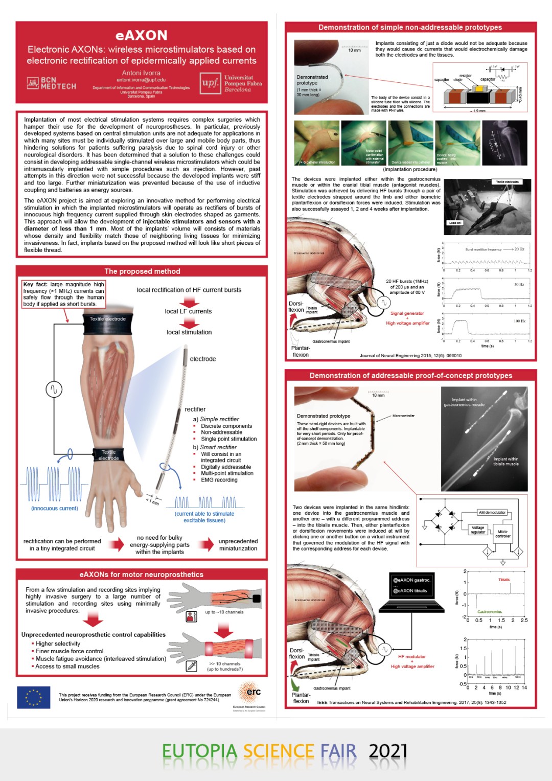
Electronic AXONs: wireless microstimulators based on electronic rectification of epidermically applied currents
ANTONI IVORRA
Department of Information and Communication Technologies Universitat Pompeu Fabra, Barcelona, SpainTo build interfaces between the electronic domain and the human nervous system is one of the most demanding challenges of nowadays engineering. Fascinating developments have already been performed such as visual cortical implants for the blind and cochlear implants for the deaf. Yet implantation of most electrical stimulation systems requires complex surgeries which hamper their use. Previously developed systems based on central stimulation units are not adequate for applications in which many sites must be individually stimulated over mobile body parts, thus hindering neuroprosthetic solutions for patients suffering paralysis due to spinal cord injury or other neurological disorders. A technological solution to address this challenge can consist in deploying a network of addressable wireless microstimulators implantable with simple procedures such as injection. Such solution was proposed and tried in the past. However, previous attempts did not achieve satisfactory success because the developed implants were stiff and too large. That is, those devices were too invasive for dense implantation. Further miniaturization was prevented because of the use of inductive coupling and batteries as energy sources. In the eAXON project we are exploring an innovative method for performing electrical stimulation in which the implanted microstimulators will operate as rectifiers of bursts of innocuous high frequency current supplied through skin electrodes shaped as garments. This approach has the potential to reduce the diameter of the implants to less than 1 mm and, more significantly, to allow that most of the implants’ volume consists of materials whose density and flexibility match those of neighbouring living tissues for minimizing invasiveness. In fact, implants based on the proposed method will look like short pieces of flexible thread.
The main objective of the eAXON project is to develop and to in vivo demonstrate electrical stimulation systems consisting of addressable wireless microelectronic implants whose actuation principle will be based on electronic rectification of epidermically applied currents. These systems will exhibit an unprecedented level of minimal invasiveness which, in turn, will yield novel modes of application of electrical stimulation for therapeutics. A subjacent objective is to develop and demonstrate these systems specifically aiming at neuromuscular stimulation for developing neuroprosthetic systems.
Another objective of the eAXON project is to illustrate that galvanic coupling through living tissues at high frequencies can be effectively and safely used for powering electronic implants in general; as an alternative to current energy transfer and harvest methods which require embedding bulky components within the implants.
- Lunder, University of Ljubljana
-
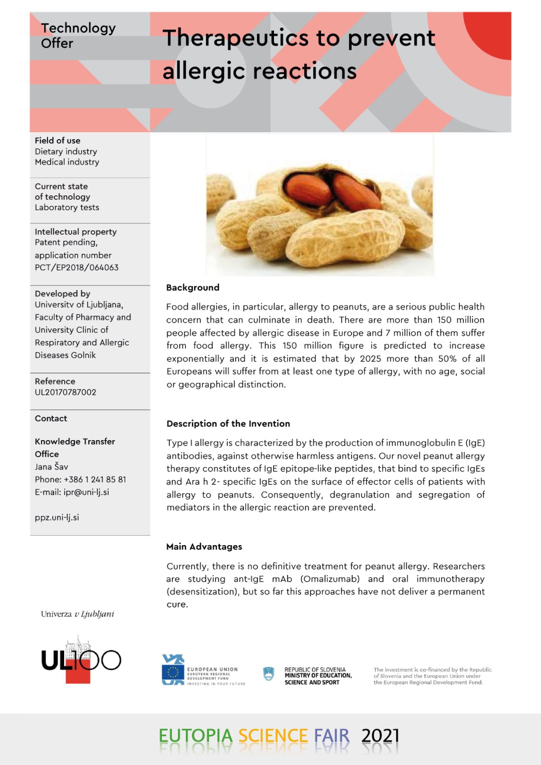
Therapeutics to prevent allergic reactions
- 2.5 The World of Life: Chemistry, Genetics, Medicine. Part 2
-
- A. Gand et al., CY Cergy Paris Université
-
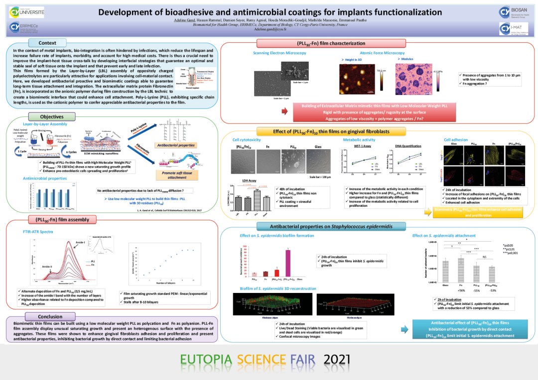
Development of bioadhesive and antimicrobial coatings for implants functionnalization
ADELINE GAND, DAMIEN SEYER, HASSAN RAMMAL, RÉMY AGNIEL, HOUDA MORACHKI-GOUDJIL, MATHILDE MASSONIE, EMMANUEL PAUTHE
Biomaterial for Health Group, ERRMECe, Department of Biology, Cergy-Paris University, France Adeline.gand@cyu.frIn the context of orthopedic/dental implants, bio-integration is often jeopardized by infections which reduce the lifespan and increase failure rate ofimplants, morbidity, and account for high medical costs. There is thus a crucial need of enhancing the implant-host tissue cross-talk by developinginterfacial strategies that guarantee an optimal and stable seal of soft tissue onto the implant and that prevent early and late infection. Thin films formed by the Layer-by-Layer (LBL) assembly of oppositely charged polyelectrolytes are particularly attractive for applications involving cell-material contact. Here we developed proactive and biomimetic coatings, able to guarantee long- term tissue attachment and integration, withantimicrobial properties. The extracellular matrix protein Fibronectin (Fn), is incorporated as the anionic polymer during film construction by the LBL technic to create a biomimetic interface that could enhance cell attachment. Poly-L-Lysine (PLL)-exhibiting specific chain lengths- is used asthe cationic polymer to confer remarkable antibacterial property to the film.
In a previous study we have shown that it was possible to build PLL-Fn thin films, using a 70-150 kDa PLL as polycation and Fn as polyanion. We observed that these films presented a novel terminated growth, and were capable of enhancing cell adhesion, spreading and proliferation compared to a Fn monolayer. Nevertheless, these thin films showed no antibacterial properties, most probably due to the high molecular weight of the PLL that prevent the polyelectrolyte to diffuse within the film. Thin films were thus constructed using a low molecular weight PLL (PLL30). These filmsshowed the same unusual terminated growth as previously described and are able to enhance the proliferation and adhesion of primary gingival fibroblasts in comparison with a Fn monolayer. Antibacterial effects were tested on 24h-old biofilm formed by S. epidermidis. The results showed a strong inhibition of bacterial growth and 3D visualization of biofilm subsequently stained with Syto-9 and PI showed that bacteria in direct contactwith film are dead. Moreover, thin films were effective in limiting initial Staphylococcus attachment (2h) with a reduction of 53%. These findings shed the light on the potential of thin films, constructed with Fn and PLL30, to enhance cell proliferation and attachment while avoiding bacterialinfection.
- Q. Marcelis, Vrije Universiteit Brussel
-
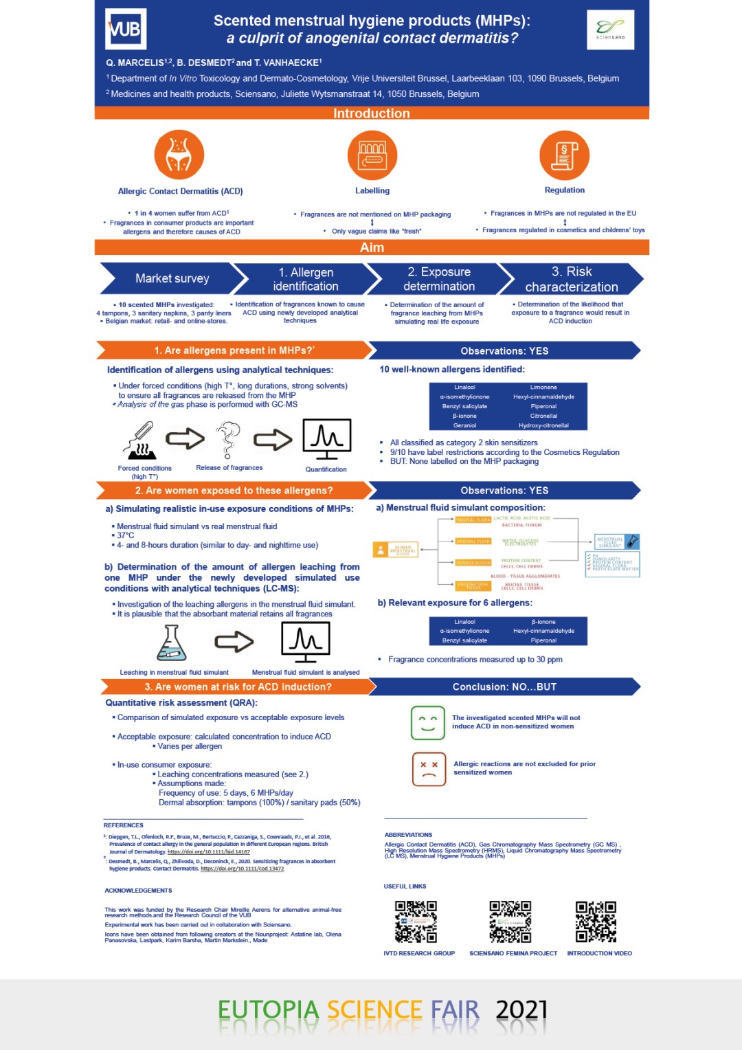
Scented menstrual hygiene products (MHPs): a culprit of anogenital contact dermatitis?
Q. MARCELIS (1,2), B. DESMEDT (2) AND T. VANHAECKE (1)
(1) Department of In Vitro Toxicology and Dermato-Cosmetology, Vrije Universiteit Brussel, Laarbeeklaan 103, 1090 Brussels, Belgium.
(2) Medicines and health products, Sciensano, Juliette Wytsmanstraat 14, 1050 Brussels, BelgiumMore than one-quarter of the general European population suffers from ACD, with a higher prevalence in women than men. Often, ACD is provoked by fragrance allergens present in scented consumer products. ACD of the anogenital region is less common but equally impairs the quality of life. To attract consumers, vaginal hygiene products such as intimate wipes and menstrual hygiene products (MHPs) are often scented with numerous fragrances. However, ingredient labeling is not mandated in the EU for menstrual hygiene products. This results in vague terms like ‘fresh’ on the packaging, but no adequate disclosure of which allergens are present in these products. Knowing that fragrance allergens are a frequent cause of ACD, combined with the less stringent regulation on ingredient disclosure and the limited barrier function of the vaginal environment, the question arises whether or not scented MHPs are safe for the consumer.
Ten scented MHPs (4 tampons, 3 sanitary napkins, 3 panty liners) originating from the Belgian market have been investigated on the risk for ACD induction. ACD induction depends on the skin sensitizing potency of the fragrance ingredient, the exposure, and the frequency of use. To investigate this risk, fragrance allergens are quantified in tampons and sanitary napkins, simulating real-life consumer exposure in a so-called leaching experiment. Our in-house developed menstrual fluid simulant allowed us to simulate this exposure accurately (2). Last, the risk of ACD development is assessed by comparing acceptable exposure levels described in the literature with the determined consumer exposure level.
In total, ten fragrance allergens have been identified in the investigated MHPs. These are well-known skin sensitizers, of which nine have restrictions laid down in the Cosmetics Regulation. Six fragrance allergens leach in quantifiable amounts from the scented MHPs when simulating the exposure. When the potency, exposure, and frequency of use are taken into consideration, it is expected that the MHPs would not induce ACD. However, the risk assessment does not take ACD elicitation into account for prior sensitized women.
Scented MHPs are a relevant source of exposure from fragrance allergens. Fragrance allergens are not indicated on the packaging of MHPs. There is no risk for ACD induction solely by MHPs use. However, ACD elicitation is still possible for prior sensitized women.
- C. Fontsere et al., University of Pompeu Fabra
-
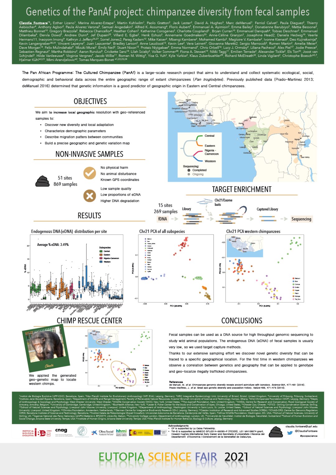
Genetics of the PanAf project: chimpanzee diversity from fecal samples
CLAUDIA FONTSERE ET AL.
University of Pompeu Fabra
Knowledge on the population history and connectivity of free-ranging endangered species is critical for understanding their conservation status and planning their management. Whole-genome data on chimpanzees (Pan troglodytes) is sparse, precluding the description of fine-grained population connectivity in space and time. To overcome this, we have produced the first non-invasive geolocalized catalogue of genomic diversity based on single nucleotide variation by capturing the complete chromosome 21 from 828 fecal samples collected at 48 sampling sites. These yield genetic evidence for the four recognized subspecies diverging during the Middle Pleistocene. Despite their long-term genetic differentiation, often correlating with known geographical barriers, we found previously undetected areas of genetic exchange, including differential admixture with bonobos within the same subspecies. The geographic distribution of samples allowed us to identify intra-subspecies population stratification, and reconstruct fine-scale patterns of isolation, migration and connectivity during the Late Pleistocene and Holocene until recent times. Our findings imply that chimpanzees, unlike humans, did not experience extended episodes of long-distance migrations. Finally, the extensive sampling revealed substantial amounts of heretofore undescribed local variation. This allowed us to generate a fine-grained geo-genetic map for the geolocalization of samples, with a good precision of on average 100 km from their true location. This genetic map of georeferenced samples could be then used as a novel tool for conservation by detecting new routes for illegal trade, with the potential to establish the first atlas of geographical origin of poached or confiscated chimpanzees. [T. Márques-Bonet] - A. Inman, University of Warwick
-
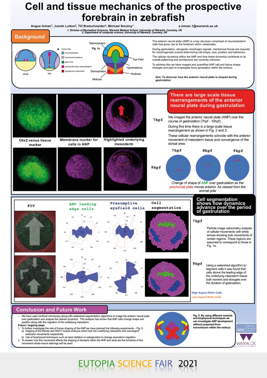
Cell and tissue mechanics of the prospective forebrain in zebrafish
ANGUS INMAN (1), JOSIAH LUTTON (2), TILL BRETSCHNEIDER (2), MICHAEL SMUTNY (1)
(1) Division of Biomedical Sciences, Warwick Medical School, University of Warwick, Coventry, UK
(2) Department of computer science, University of Warwick, Coventry, UKBackground
a.inman.1@warwick.ac.ukThe anterior neural plate (ANP) is a key structure in vertebrates, comprised of neuroectoderm cells, giving rise to the early forebrain. Along with morphogenic signals required for patterning of the tissue, mechanical forces are required for various morphogenetic events and required to determine cell shape, size, position and behaviour. This patterning process becomes apparent during gastrulation with the formation of distinct functional domains containing similarly fated cells. The role of mechanical forces during this process of ANP morphogenesis and patterning is currently poorly understood is the focus of my project.
Using the zebrafish as a model organism ANP cell and tissue shape changes can be imaged and quantified allowing us to probe the tissue mechanics of the ANP. Cell positions and shapes will then be tracked permitting inferences of the distribution and magnitude of forces within the tissue. The role of myosin will be visualised using immunostainings and adapted using an optogenetic setup. Mutant embryos will also be used to give us insight into how global morphogenetic movements and external tissues may have an effect on the neural plate.
We are also using micorluidic devices to study the mechanical properties of individual cells, creating an extensional flow that will deform cells and thus highlight any differences in reactions between cell populations.
- L. Garbutt, University of Warwick
-
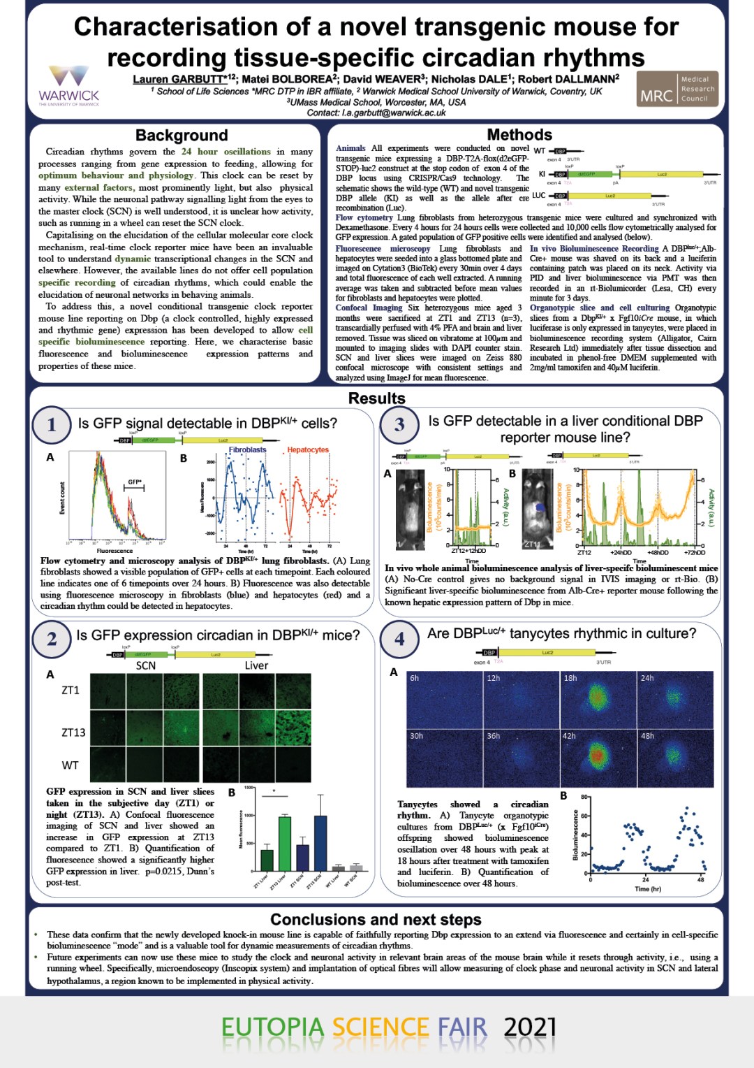
Characterisation of a novel transgenic reporter mouse for recording tissue-specific circadian rhythms
LAUREN GARBUTT (1,2), MATEI BOLBOREA (3), DAVID R. WEAVER (4) AND ROBERT DALLMANN (3)
(1) School of Life Sciences,
(2) MRC DTP in IBR,
(3) Warwick Medical School, University of Warwick, UK.
(4) Department of Neurobiology, University of Massachusetts Medical School, Worcester, MAThe discovery of the genes involved in the molecular circadian clock and its outputs has led to a deeper understanding of the functional importance of circadian clocks in many processes. One key technique to track the molecular clock in real-time is the use of reporter systems such as PER2::LUC, as well as other circadian reporter mice which have been invaluable to drive discoveries in the field. However, due to the ubiquitous expression of luciferase in these mouse models, measurement of circadian rhythms, from specific cell types of heterogenous tissues to organ specific expression in vivo, is limited.
Here, we describe the novel, transgenic mouse line DBP-T2A-flox(d2eGFP-STOP)-Luc2, in which the clock-controlled gene dbp elicits the expression of GFP (DBPKI+), and following cre recombination, is capable of displaying cell-type specific bioluminescence (DBPLuc/+).
Confocal fluorescence microscopy of DBPKI+ livers and SCN ex vivo showed a clear oscillation in GFP expression over 24 hours. Bioluminescence studies also showed a clear oscillation following cell-specific CRE mediated recombination in a number of cell types including liver in vivo and tanycytes ex vivo.
While further experiments are warranted to show that insertion of the reporter construct into the DBP locus does not affect DBP transcriptional activity, preliminary data suggests that these mice are a useful tool to measure circadian rhythms in specific cell types in real-time, both in vitro and in vivo using bioluminescence.


